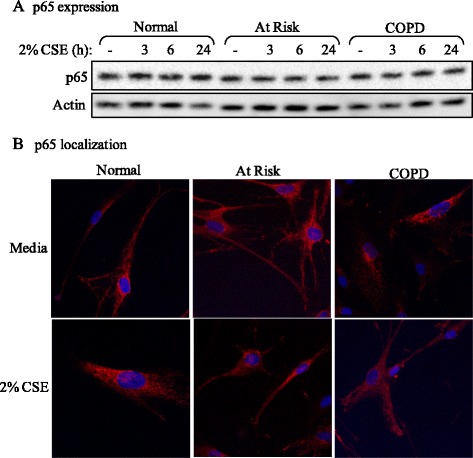
Figure 6.

p65 expression and localization in Normal, At Risk and COPD lung fibroblasts. (A) p65 expression: There was no perceptible difference in p65 expression between Normal, At Risk and COPD-derived lung fibroblasts exposed to 2% CSE for up to 24 hours. Representative western blot is shown of 3 independent experiments. (B) p65 localization: There was little perceptible difference in the localization of p65 in lung fibroblasts from the three subject groups, which appeared predominantly cytoplasmic (red colour). Exposure to 2% CSE for 30 minutes did not appreciably alter the localization of p65. Representative images shown are based on two independent experiments.
