Figure 1.

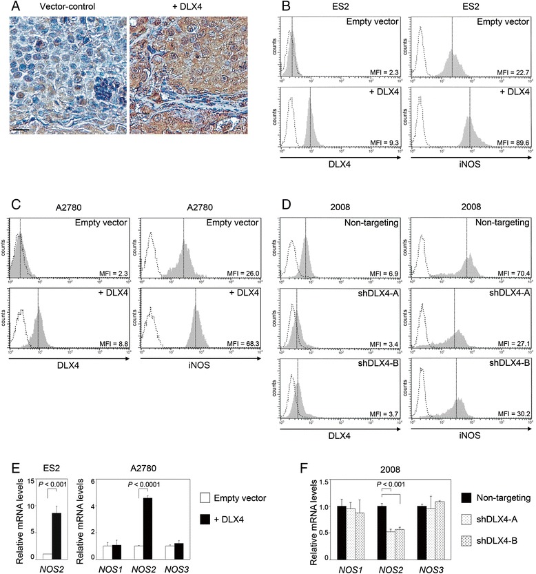
DLX4 induces iNOS expression. (A) Staining of iNOS in sections of peritoneal tumors of mice that were inoculated with vector-control and +DLX4 ES2 lines. Bar, 20 μm. (B, C and D) Flow cytometric analysis of intracellular staining of DLX4 and iNOS in transfected ovarian cancer cell lines. Mean fluorescence intensities (MFI) of staining are indicated. Shown are representative examples of DLX4 and iNOS staining in (B) vector-control and +DLX4 ES2 cells, (C) vector-control and +DLX4 A2780 cells and (D) 2008 cells transfected with non-targeting shRNA and shRNAs that targeted two different regions of DLX4 (shDLX4-A, shDLX4-B). (E) qRT-PCR analysis of relative NOS2 mRNA levels in ES2 cells and NOS1, NOS2 and NOS3 mRNA levels in A2780 cells. Levels of each mRNA in +DLX4 cells are expressed relative to the level in vector-control cells. (F) qRT-PCR analysis of relative NOS1, NOS2 and NOS3 mRNA levels in 2008 cells. Levels of each mRNA in DLX4 shRNA-transfected cells are expressed relative to the level in non-targeting shRNA-transfected cells.
