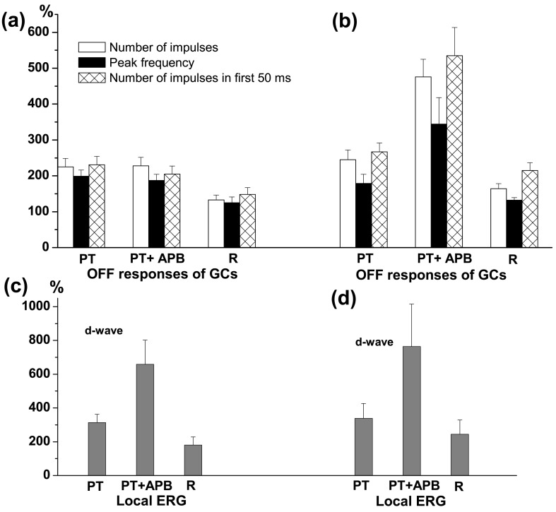
Figure 3.
Effects of perfusion with picrotoxin (PT), PT+APB and Ringer solution in the recovery period (R) on the OFF responses of ganglion cells and d-wave in local ERG. (a) Changes of mean number of impulses (white columns), peak frequency (black columns) and number of impulses in the first 50 ms (hatched columns) of the OFF responses of ON-OFF and phasic OFF GCs expressed as % from their initial values, obtained in cells with blocked enhancing effect of APB on their OFF responses during the perfusion with PT+APB. The mean ± S.E.M. are represented. (b) Changes of the same parameters of the GCs’ OFF responses as (a), obtained in cells with preserved enhancing effect of APB on their OFF responses during the perfusion with PT+APB. (c) and (d): Amplitude of the d-wave of the local ERG (mean ± S.E.M.), expressed as % from its initial value during perfusion with PT, PT+APB and Ringer (during recovery period), recorded simultaneously with activity of GCs. It is seen that the enhancing effect of APB on the d-wave amplitude is preserved during the GABAergic blockade in all eyes irrespective of where the perfusion with PT+APB prevents (c) or does not change (d) the effect of APB on the ganglion cell OFF responses.

