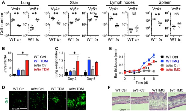
A Absolute numbers (per mouse) of indicated γδT-cell subsets in lung, skin, lymph nodes (inguinal, axillary, and submandibular), and spleen from 5-week-old WT or tn/tn mice (n = 3). Each circle represents an individual mouse, and horizontal bars indicate the mean. The statistical significance was calculated with an unpaired one-tailed Student's t-test. *P < 0.05; **P < 0.01; NS, not significant.
B–D WT or tn/tn mice were injected intravenously with an oil-in-water emulsion containing TDM (15 μg). Emulsion without TDM was injected as a vehicle control. Il17amRNA levels in lungs at day 2 were examined by quantitative RT–PCR and normalized to GapdhmRNA (n = 3–4, mean ± SEM) (B). Lung inflammation intensity was measured by calculating the lung weight index (n = 3–5, mean ± SEM) (C). The statistical significance between TDM-treated WT and tn/tn mice was calculated with an unpaired one-tailed Student's t-test. *P < 0.05. Lung sections at day 2 were stained for granulocyte marker Gr1 (D).
E, F WT or tn/tn mice were treated daily for 6 days with IMQ cream or control cream on the ears. Ear skin thickness at the days indicated (n = 3, mean ± SEM) (E). The statistical significance between IMQ-treated WT and tn/tn mice was calculated with an unpaired one-tailed Student's t-test. *P < 0.05; **P < 0.01. HE staining of the ear section at day 6 (F).