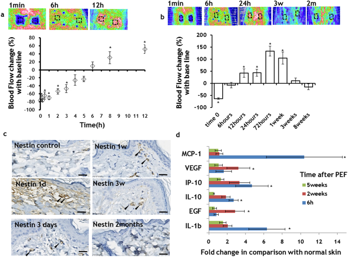
Figure 4. Controllable angiogenesis induction in the rat skin with PEF.
a PEF led to a temporary vasoconstriction at the treated site. The flow returned to the baseline levels 5 hours after PEF application. In the following hours, we observed increase of the flow at the PEF treated areas. b The increased flow almost returned to the baseline levels three weeks after PEF treatment. c Angiogenesis marker Nestin immunohistochemistry. Striking increase in the Nestin expression in the papillary dermis capillaries (black arrows) in comparison to untreated skin was observed from one day to three weeks after PEF. The intensity of staining two months after PEF was very similar to control, suggesting the maturation of the vessels. a-c Five animals were used for each time point. Error bars show ± SEM. d Secretion of pro-angiogenesis factors to the PEF treated area of the skin. All samples were normalized to the total protein. The bars show fold increase at the treated areas in comparison to expression levels in untreated animals. The bars show the average from measurements between 6 treated areas in 3 different animals. Error bars show ± SEM. (*) p-val < 0.05. Statistical analysis was performed by first using one-way ANOVA for multivariate analysis and post hoc using Dunnett’s tests to assess significance between individual groups. Significance was set at P < 0.05.

