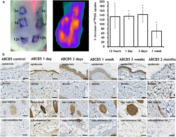
Figure 5. Increased skin metabolism after PEF administration.
The increased metabolism of skin areas treated by PEF was detected using both a 18FDG uptake (Top panel) and b Increased expression of ABCB5, between 1 day to 3 weeks after PEF treatment. The increased expression of ABCB5 was observed in subpopulations of keratinocytes, cells in the dermis, cells in the hair follicles (in various parts of the follicle) and in subcutaneous fat. For 18FDG measurements, three rats were used with two treated areas per time point. Error bars show ± SEM. For ABCB5 expression, 5 rats were used for each time point. (*) p-val < 0.001. Statistical analysis was performed by first using one-way ANOVA for multivariate analysis and post hoc using Dunnett’s tests to assess significance between individual groups. Significance was set at P < 0.05.

