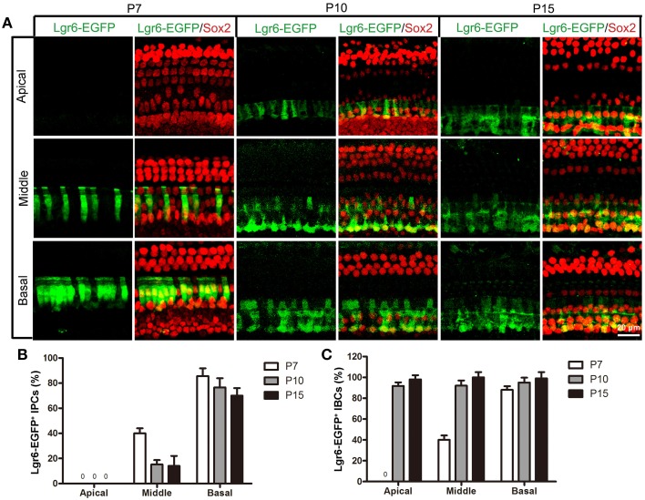
Figure 5.
Lgr6-EGFP expression decreased in the IPCs and increased in the IBCs from P7 to P15. (A) From P7 to P15, Lgr6-EGFP was expressed in both the IPCs and IBCs in the middle and basal turns. In the apical turn, there was no Lgr6-EGFP expression at P7, and Lgr6-EGFP was only expressed in the IBCs at P10 and P15. (B) From P7 to P15, the percentage of Lgr6-EGFP-positive IPCs gradually decreased (n = 4), (C) From P7 to P15, the percentage of Lgr6-EGFP-positive IBCs gradually increased (n = 4). IPCs, inner pillar cells; IBCs, inner border cells.

