
Figure 1.
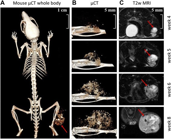
Representative MRI and μCT images of mouse xenografts 4 to 8 weeks after intratibial transplantation of patient-derived human OS tissue showing tibial tumor mass. A): μCT image of whole mouse body 8 weeks after tumor inoculation. B) and C): μCT and T2 weighed (T2w) MRI images of tumor growth from week 4 to week 8. Red arrows indicate tumor location.