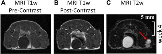
Figure 2.

Representative MRI images of T1 weighed (T1w) and T2 weighed (T2w) performed at week 4. A) and B): In T1w MRI images pre and post contrast tumor is not visible. C): Using T2w MRI imaging leasion size of 48 μl (here equaling tumor size) can be discriminated (red arrow).
