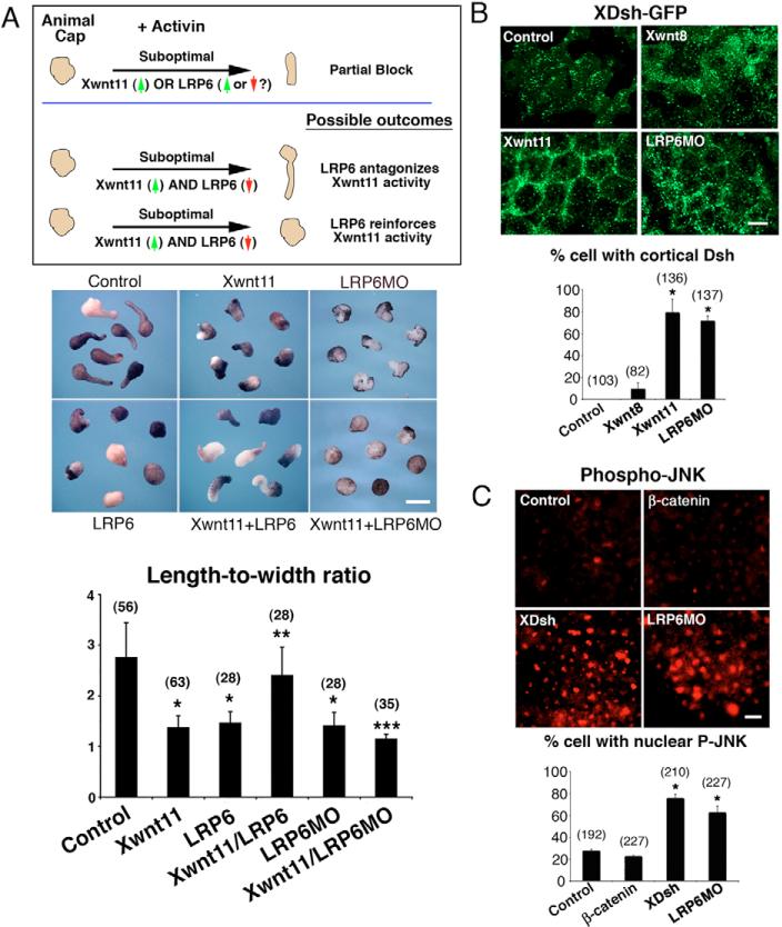
Fig. 5. Lrp6 antagonizes Wnt/PCP signaling upstream of Dishevelled and JNK.
(A) Injection of suboptimal amounts of wnt11 mRNA (Xwnt; 160 pg), an activator of Wnt/PCP signaling, or suboptimal amounts of lrp6 mRNA (LRP6; 1 ng) causes incomplete block of activin-induced animal cap elongation. Injection of suboptimal lrp6 mRNA (1 ng) reverses the partial block by Wnt11 (160 pg) of activin-mediated animal cap elongation, suggesting that Lrp6 opposes Wnt11-mediated activation of the Wnt/PCP pathway. Injection of suboptimal Lrp6MO (30 ng) reinforces the Wnt11-mediated partial block of activin-treated animal caps, further demonstrating that Lrp6 antagonizes Wnt/PCP signaling. Double asterisks for Wnt11+Lrp6 (Xwnt11/LRP6) indicate statistically significant differences from Wnt11 and Lrp6 (P<0.01). Triple asterisks for Wnt11+Lrp6MO mark differences statistically significant from Wnt11 and Lrp6MO (P<0.01). (B) Dsh-GFP translocates to the cell cortex upon Wnt/PCP activation. Injection of Wnt11 (400 pg) or Lrp6MO (40 ng) promotes cortical translocation of Dsh-GFP. By contrast, injection of the Wnt/β-catenin ligand, Wnt8 (Xwnt; 40 pg), has no significant (bar chart) effect on XDsh-GFP localization. (C) Jun-N-terminal kinase (JNK) is phosphorylated and localized to the nucleus in animal caps injected with dsh (XDsh; 1 ng) (160 pg) or Lrp6MO (40 ng). Animal caps injected with β-catenin mRNA (50 pg) demonstrate minimal phospho-JNK staining or nuclear localization similar to uninjected control. Student's t-test was used for statistical analysis. Error bars indicate standard deviation. Asterisks mark differences that are statistically significant from control (P<0.01). Numbers of explants/cells scored are indicated in parentheses. Scale bars: 500 m in A; 50 μm in B; 100 μm in C.

