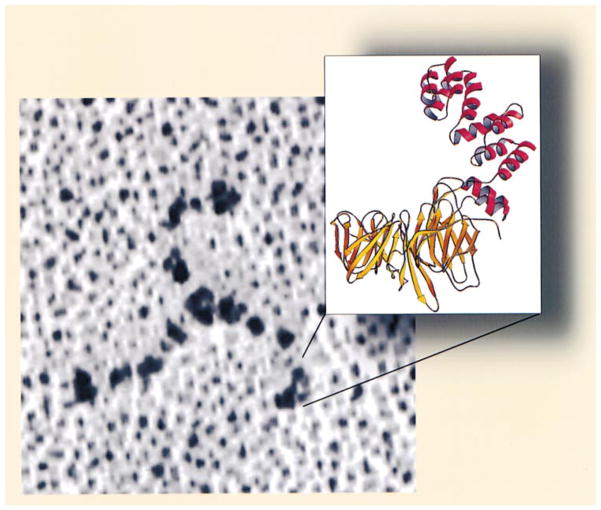
Figure 8. Relationship between the Clathrin Triskelion and the Structure of the Terminal Domain and Linker.
Clathrin was deposited onto freshly cleaved mica by spraying in 50% glycerol, rotary shadowed with Pt, and visualized by electron microscopy (see Heuser and Kirchhausen, 1985). The ribbon diagram corresponds to the terminal domain and linker.

