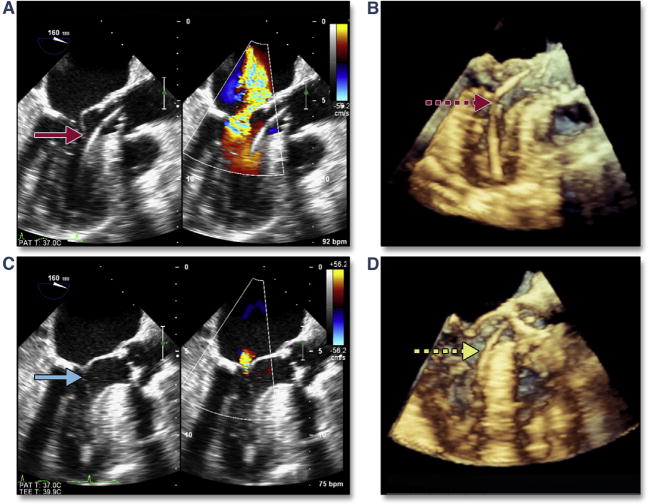
Figure 1. Acute Severe Mitral Regurgitation.

During stiff wire positioning, malcoaptation of the mitral valve leaflet (A, red arrow) (Online Video 1), resulting in severe mitral regurgitation, may be the first clue to entanglement of the wire in the mitral apparatus (B, dashed red arrow). With repositioning of the wire, coaptation of the mitral valve is now normal (C, blue arrow) with mild mitral regurgitation. The correct position of the wire is confirmed by using 3-dimensional imaging (D, dashed yellow arrow).
