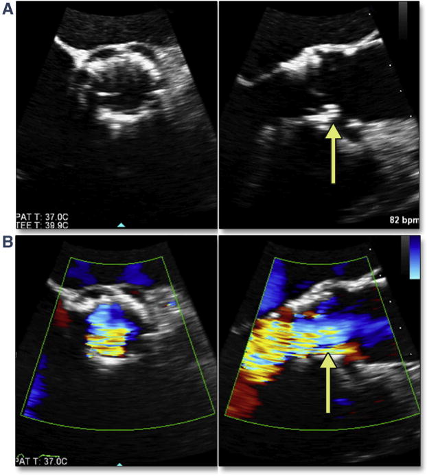
Figure 11. Malpositioning of the THV.

Positioning of the THV too ventricular (low), with failure to cover the native cusps, risks native leaflet overhang. In this simultaneous multiplane image, the THV leaflet has become entrapped by the bulky calcium of the overhanging native cusp (A, yellow arrow) (Online Video 10), resulting in significant aortic regurgitaton (B, yellow arrow) (Online Video 11). Abbreviation as in Figure 8.
