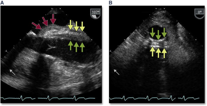
Figure 17. Intramural Hematoma.

Bleeding into the wall of the aorta is seen as thickening of the wall between the adventitia (yellow arrows) and endothelium (green arrows). In the long axis view (A), this may be associated with periaortic hematoma (red arrows). (B) shows extension into the descending aorta with a circumferential thickening of the wall.
