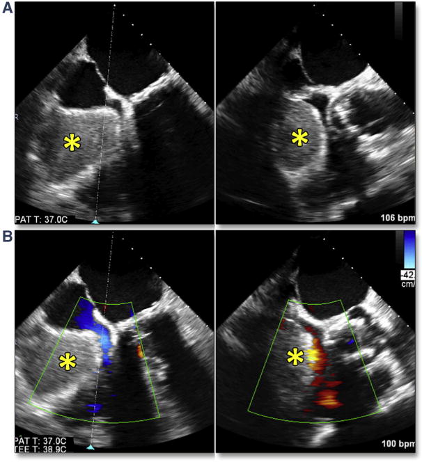
Figure 22. Acute Right Ventricular Perforation.

After successful TAVR with no evidence of conduction abnormalities, the pacing wire was removed with immediate accumulation of blood seen by simultaneous multiplane imaging within the pericardial space (A) (Online Video 23) adjacent to the right ventricle (*). This resulted in obstruction to the tricuspid valve flow (B) (Online Video 24) and ensuing tamponade physiology. Abbreviation as in Figure 5.
