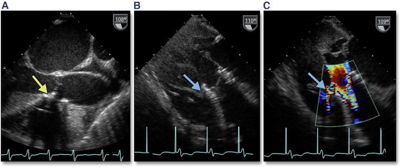
Figure 25. Ventricular Septal Rupture.

Ectopic calcification may extend into the LVOT along the membranous septum (A, yellow arrow). After TAVR, deep gastric long axis views may be the best views for imaging the defect in the membranous septum (B, blue arrow) (Online Video 27) with color Doppler (C) (Online Video 28) showing systolic flow across a traumatic ventricular septal defect. Abbreviations as in Figures 5 and 7.
