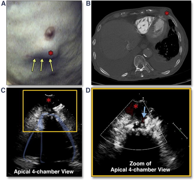
Figure 33. Apical Pseudoaneurysm Formation After Transapical Cannulation.

This patient complained of an expanding chest wall mass (A, red asterisk) 6 months after transapical TAVR (yellow arrows show the mini-thoracotomy scar). Transthoracic imaging revealed a pseudoaneurysm, best seen from the apical views (C, red asterisk) (Online Video 39) with systolic flow detected on color Doppler (D, blue arrow) (Online Video 40). The finding was confirmed by both echocardiographic intravenous contrast study as well as chest computed tomography (CT) (B). Abbreviation as in Figure 5.
