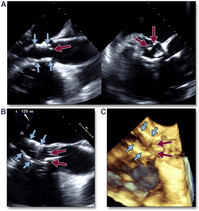
Figure 35. Translocation of the THV into the Ventricle.

Migration of the THV into the LV can occur when the initial position of the valve is too low. The simultaneous multiplane image (A) (Online Video 41), with the long-axis view of the transcatheter valve on the left and the short-axis view on the right, shows the low position of the transcatheter valve (blue arrows) with significant native leaflet overhang (red arrows). Translocation of the transcatheter valve is seen by both 2-dimensional (B) and 3-dimensional (C) (Online Video 42) imaging. Abbreviations as in Figures 8 and 30.
