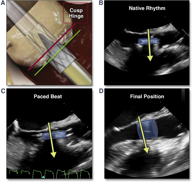
Figure 9. Positioning of the THV.

(A) (modified from Dvir et al. [67]) shows the desired fluoroscopic position of the valve just before balloon inflation (during rapid pacing). (B) shows the transesophageal echocardiographic position during native rhythm and in diastole, which results in a superior positioning during pacing (C). Shortening of the valve results in positioning of the proximal edge of the THV 1 to 2 mm below the native annulus (D) (Online Video 9). Abbreviation as in Figure 8.
