Figure 1.

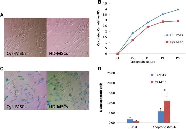
Biological characterization of HD- and Cys-MSCs. A: Morphology of bone marrow (BM)-derived mesenchymal stromal cells (MSCs) isolated from the cystinotic patient (Cys-MSCs, on the left) and from one representative healthy donor (HD-MSCs, on the right) at passage 2. Magnification x10. Cys-MSCs display the typical spindle-shape morphology of HD-MSCs. B: Calculated population doublings (PDs) from P1 to P5 of HD-MSCs (mean of 3 different HDs) and Cys-MSCs. Cys-MSCs show a lower proliferative capacity, as compared with HD-MSCs. C: ß-Galactosidase staining of MSCs isolated from the cystinotic patient (Cys-MSCs on the left) and from one representative healthy donor (HD-MSCs on the right). Senescence of the cells is indicated by the positivity for the staining. D: After cysteamine treatment, the percentage of Cys-MSCs late apoptotic cells becomes significantly higher than HD-MSCs’ one.*Indicates a p value lower than 0.05.
