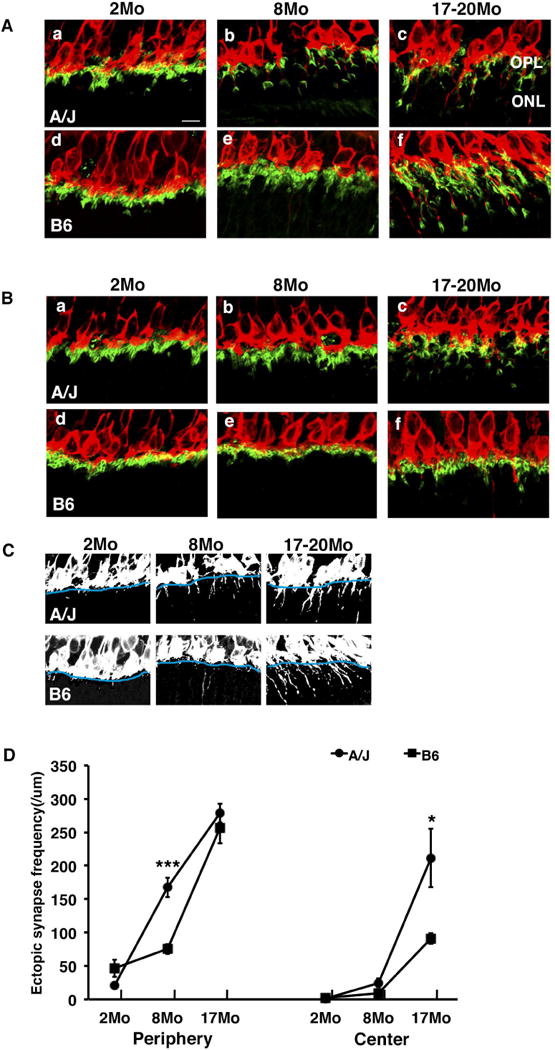
Fig. 1.

Ectopic synapse localization in A/J and B6 mice. (A) In the peripheral retina, PKCα-labeled (red) bipolar cell dendrites and PSD95-labeled (green) photoreceptor synaptic terminals are localized in the OPL in B6 mice at 2 and 8 months of age (d, e) and in A/J mice at 2 months of age (a). They are ectopically localized in the ONL in A/J mice at 8 and 17–20 months (c) and in B6 mice at 17–20 months of age (f). (B) In the central retina, the synaptic terminals are localized to the OPL in B6 mice at 2 and 8 months of age (d, e). At 17–20 months of age B6 mice exhibit some ectopically localized synapses (f), but it is significantly less than A/J mice. A/J mice exhibit normal localization in OPL at 2 and 8 months (a, b) but ectopically localized synapses in ONL at 17–20 months (c). (C) Black and white images of a–f in figure 1A demonstrate the method used for quantification of ectopic synapses. The OPL boundary is identified in blue, and the number of synapses extending past the boundary was counted. (D) A/J mice exhibit a significant increase in ectopically localized synapses in the peripheral retina at 8 months of age compared to B6 mice (left). At 17–20 months, A/J mice exhibit a significant increase in ectopic synapses in the central retina compared to B6 mice (right). Error bars represent standard errors. Asterisks denote statistical significance (*P < 0.05, **P < 0.01, ***P < 0.001). OPL, outer plexiform layer; ONL, outer nuclear layer; scale bar, 10 um.
