Fig. 2.

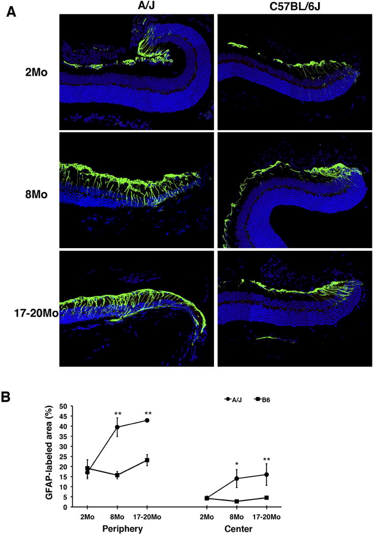
Retinal stress marker in A/J and B6 mice. (A) In the peripheral B6 retina, GFAP (green), a marker of retinal stress, exhibits staining that is consistent at all timepoints examined (right). In A/J mice, GFAP staining becomes higher in intensity and extends further towards the center of the retina as mice age (left). (B) In both the peripheral (left) and central (right) retina, the percentage of GFAP-labeled area of the retina is significantly higher in A/J mice compared to age matched B6 mice at 8 and 17–20 months. Asterisks denote statistical significance (*P < 0.05, **P < 0.01, ***P < 0.001).
