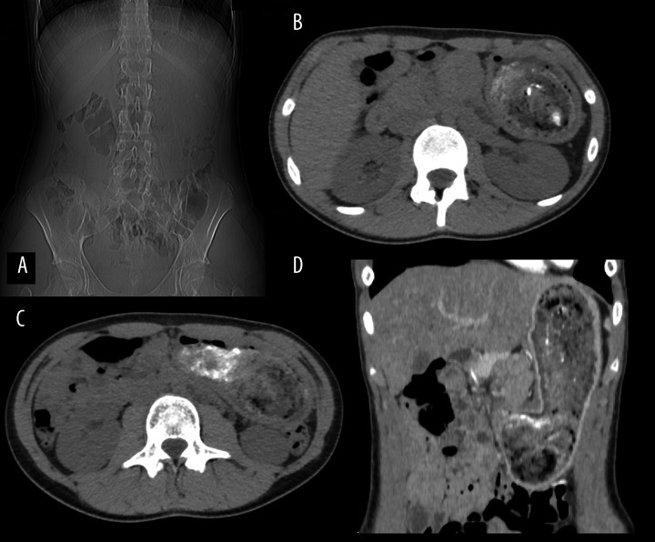
Figure 8.
Female patient aged 13 – invisible gas in the stomach in the pilot sequence of CT (A); a mixture of hair and hyperdense plaster in native scans (B, C); coronal reformatted CT scan after intravenous injection of the contrast medium – stretched stomach containing a non-enhancing mass separated from the stomach wall (D).

