
Fig. 6.
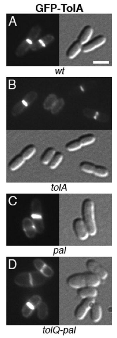
Localization of GFP–TolA to the division site in wt and mutant cells. GFP–TolA in wt (A), tolA (B), pal (C) and tolQ-pal (D) cells. Note the normal morphology of cells in B. Strains used were TB28 (A), FB20229 (B), MG5 (C) and MG4 (D), each harbouring pNP4 [Plac∷gfp-tolA]. Cells were grown at 30°C in M9-glucose medium supplemented with 5 μM IPTG. Bar equals 2 μm.