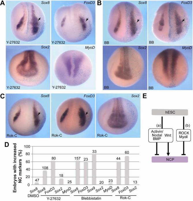
Figure 6. Inhibition of ROCK and Myosin II expands neural crest territory in Xenopus embryos.
(A-C) Albino embryos were injected unilaterally with Y-27632 (A), Blebbistatin (BB, B) or Rok-C RNA (C) at the 4-8-cell stage, cultured until stages 14-16. The neural crest genes Sox8 andFoxD3, the pan-neural marker Sox2 and the somitic marker MyoD were analyzed by in situ hybridization as indicated. Dorso-anterior views of representative embryos are shown. Arrows point to altered gene expression on the injected side, which is marked by β-galactosidase staining (light blue). (D) Quantification of the effect shown in (A-C) as percentage of embryos with increased marker expression. Total number of embryos pooled from several independent experiments is indicated at the top of each bar. (E) Pathways that convert hESC into NCPs. (a) Simultaneous inhibition of Activin/Nodal/BMP and upregulation of the Wnt pathway promotes neural crest development. (b) Inhibition of ROCK or Myosin II is sufficient to trigger NCP formation.

