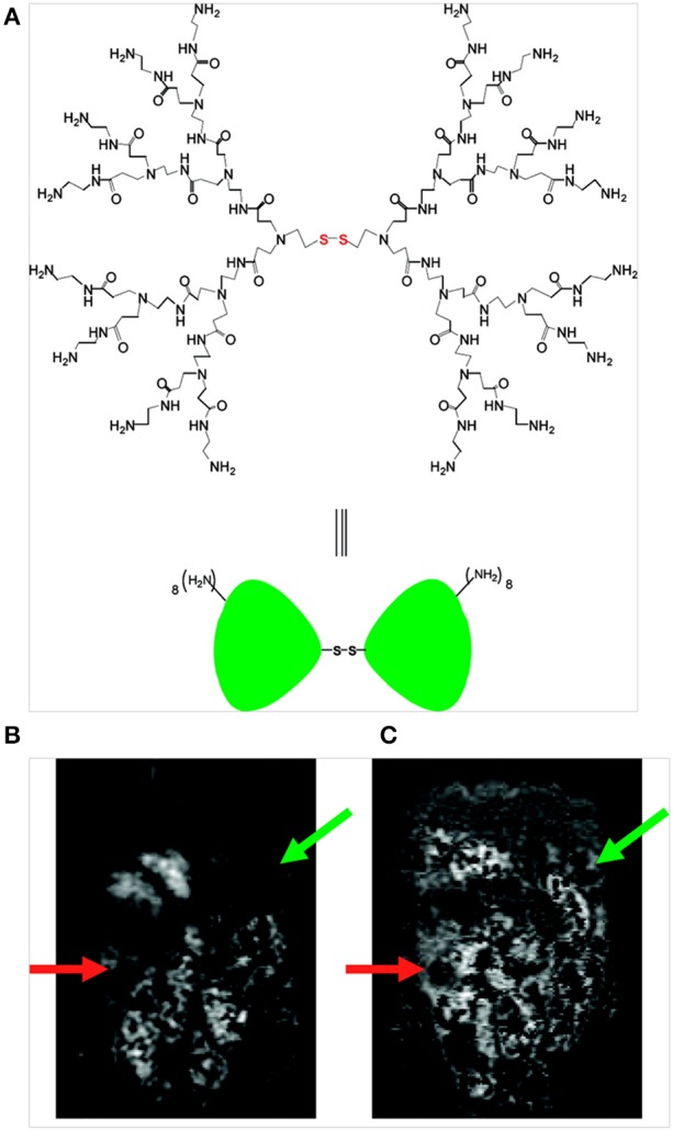
Figure 3.

(A) Structure of a PAMAM dendrimer possessing a disulfide cystamine core; (B) T1-weighed MR image obtained prior to contrast agent injection (t = 0) reveals an area of tumor growth below the liver, stomach and spleen in the left abdomen (green arrow) and adjacent to the gastrointestinal tract in the lower right abdomen (orange arrow) and (C) T1-weighted MR image obtained 6.5 h after intra-peritoneal injection of the dual contrast agent (green and orange arrows indicate signal enhancement on the tumor surface) (Xu et al., 2007), © 2007, American Chemical Society.
