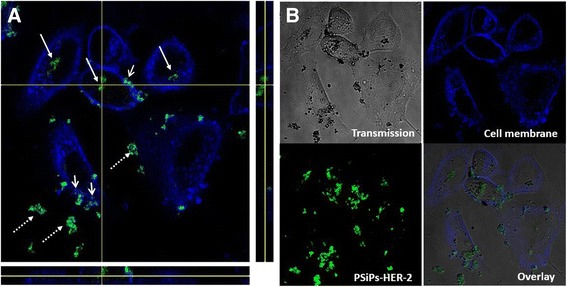
Figure 3.

Particle localization into SK-BR-3 cell lines. SK-BR-3 (cell membrane labeled in blue) treated with PSiP-HER-2 particles (presented in green) for 24 h, showing the particles inside (arrow) and, outside (dashed arrow) the cells and at the cell membrane (opened arrow). (A) Orthogonal view in the 3 planes (X/Y, X/Z and Y/Z) of the particles pointed at the intersection of the X and Y axeis. As seen from all planes, the particles are surrounded by the cell membrane. (B) Confocal images of the cells in the transmission channel, the cell membrane and PSiP-HER-2 in fluorescent channels, and overlay of all channels. PSiP-HER-2 were labeled with DyLight 488.
