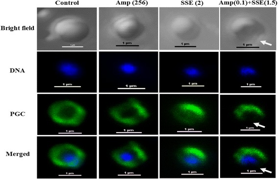
Figure 4.

Schematic representation of the results from immunofluorescence and a confocal laser scanning microscope; Samples of ARSA after treatment for 4 h with Ampicillin, SSE, either alone or in combination. Amp (256), ampicillin at 256 μg/ml; SSE (2), Stephania suberosa extract at 2 mg/ml; Amp (0.1) + SSE (1.5) = Ampicillin 0.11 μg/ml plus SSE 1.5 mg/ml. The cells were stained for DNA with DAPI (blue) and labeled for peptidoglycan (PGC) (green) using respective antibodies. DNA in all groups was localized in the central of cell and surrounded by a peptidoglycan layer (merged images). The white arrows showed explicit disruption of peptidoglycan. Scale bar = 1 μm.
