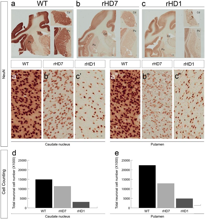
Fig 5. Reduced striatal neurons in the caudate nucleus and putamen of HD monkeys.
Immunostaining with the neuronal-specific marker NeuN in the striatum in 5-year-old control (a), rHD7 (b), and rHD1 (c) monkeys (scale bar = 1mm). The close-up view at the right side shows a closer view of the caudate nucleus and putamen (scale bar = 100μm). The number of NeuN-positive neurons in the caudate nucleus (a’, b’, c’) and putamen (a”, b”, c”) of the HD monkeys is dramatically reduced compared with the control animal (scale bar = 10 μm). By counting from the Nissl staining, the number of neurons in the caudate nucleus and putamen is reduced in both HD monkeys when compared to control monkeys (d, e, respectively).

