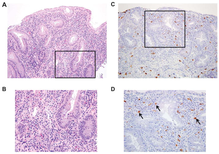
FIGURE 2.
MCs in the colonic mucosa of a patient with IBD. A patient with active UC underwent colonoscopy to determine the extent and severity of the colitis. H&E histochemistry, performed on sections of a biopsy from an inflamed segment, revealed acute and chronic inflammatory changes in the mucosa at low ×20 (A) and higher ×40 power (B). To visualize the Kit+ MCs in the biopsy, replicate sections were stained with an antibody that recognizes this tyrosine kinase receptor. Kit+ MCs were interspersed throughout the lamina propria (black arrows) at low ×20 (C) and higher ×40 power (D). There were no aggregates or sheets of MCs that would be consistent with systemic mastocytosis. Images courtesy of Jason Hornick, MD, Department of Pathology, Brigham and Women’s Hospital.

