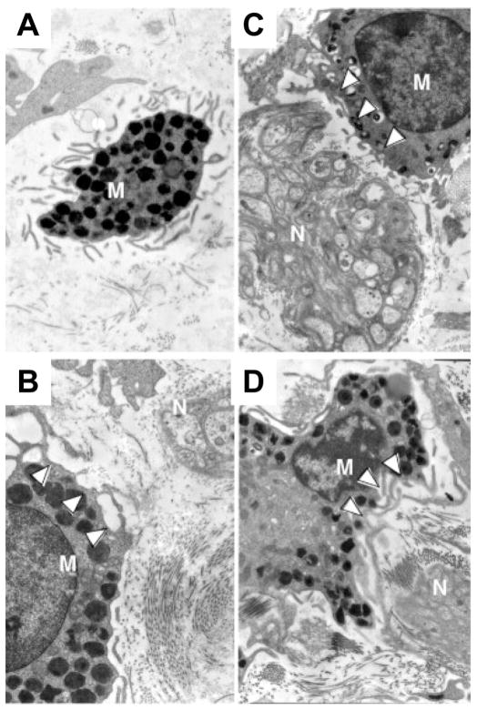
FIGURE 3.
Ultrastructure of intestinal MCs in proximity to nerve endings in GI disorders. MCs are granulocytes that can be identified in tissue section by electron microscopy based on their characteristic monolobed nucleus, elongated surface folds (microplicae), and abundant cytoplasmic electron-dense granules (A). In the intestine, MCs (M) frequently reside in close proximity to blood vessels and nerves (N). Depicted are MCs adjacent to nerve endings and in various stages of activation (B–D) in a patient with IBS. White arrowheads highlight granules that are partially or completely empty. Reprinted from Giovanni Barbara et al, Gastroenterology 2007;132:p 30 with permission from Elsevier.

