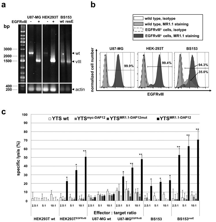
Fig. 2. Expression of EGFRvIII on target cell lines and specific cytotoxicity of MR1.1-DAP12 CAR-expressing YTS-NK cells against EGFRvIII+ tumor cells.
a: Agarose gel electrophoresis showing RT-PCR products using primers specific for EGFRvIII as well as EGFR wild type. The PCR reaction favored amplification of EGFRvIII (vIII) with approximately 1.4 kb. Note that also bands representing 2.5 kb EGFR (wt) are detected in U87-MG and HEK293T cells. As internal control an RT-PCR for actin is included. “-“ indicates wild type cells and “+” depicts cells transduced with lentiviral EGFRvIII expression vector. wt, wild type; resE, erlotinib resistant cells. b: FACS assisted analysis of EGFRvIII surface expression of U87-MGEGFRvIII, HEK293TEGFRvIII and BS153resE cells (dark grey histograms) and isogenic U87-MG and HEK293T as well as BS153 cells (light grey histograms). Staining was accomplished using c-myc-tagged scFv(MR1.1) recombinant single chain antibody. Isotype stainings using a prostate stem cell antigen (PSCA) specific scFv(AM1) are included for each isogenic cell line (open histograms). The scFvs were visualized by secondary biotin-labeled c-myc-tag specific antibody and a tertiary anti-biotin-PE antibody. Note the strong surface expression of EGFRvIII in the U87-MGEGFRvIII and HEK293TEGFRvIII target cell lines. Interestingly, BS153 parental cells contain a notable fraction of EGFRvIII-positive cells (see lower arrow) whereas BS153resE cells show a marked increase of the EGFRvIII-positive cell fraction after erlotinib treatment (94 %). c: Gene-engineered and parental YTS (wt) cells were co-cultured with 51chromuim-loaded EGFRvIII+ tumor cells and isogenic EGFRvIII- tumor control cells at different effector to target ratios for 21 h. The mean of specific tumor cell lysis and standard deviation of triplets of three chromium release assays is shown. Note the strong tumor cell lysis mediated by YTSMR1.1-DAP12 cells. (*p < 0.05, **p < 0.01).

