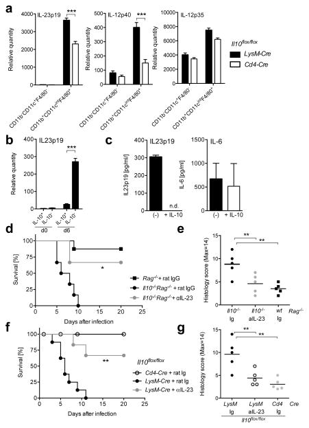
Figure 4. Macrophage-derived IL-10 influences mortality through regulation of IL-23p19.
(a) Analysis of gene expression by RT-PCR in sorted CD11b−CD11c+F4/80− DC or CD11b+CD11c+F4/80+ macrophages from large intestines in Il10flox/floxLysM- Cre or Il10flox/floxCd4-Cre mice at day 6 after C. rodentium infection. Averages and SD from two independent experiments with 6 mice in each group are shown. (b) Analysis of Il23p19 mRNA transcripts by RT-PCR in large intestine lamina propria macrophages, including GFP+(IL-10+) and GFP−(IL-10−), sorted from five to eight Il10gfp mice before and after 6 days after C. rodentium infection. These data are from 2 independent experiments. (c) Large intestine lamina propria macrophages were sorted from five to eight infected Il10−/−Rag−/− mice and cultured for 24h in the absence (black bars) or presence (white bars) of 100ng/ml recombinant mouse IL-10. Supernatants were analyzed for IL23p19 and IL-6 by ELISA. These data are from 2–3 independent experiments. (d,e) Il10−/−Rag−/− and Rag−/− mice or (f,g) Il10flox/floxLysM-Cre and Il10flox/floxCd4-Cre mice were infected with C. rodentium. (d–g) At the time of infection, 100μg anti-IL-23 or rat IgG were injected per mouse intravenously. (d, f) Survival curve and (e,g) histology score from middle colon at day 6 after infection are shown. These are from 2 independent experiments with 3–4 mice per group. Student’s t test, * p<.05, ** p<.01, *** p<.001.

