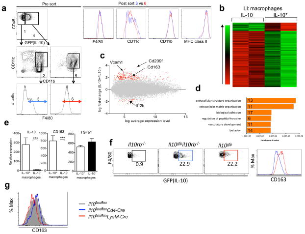
Figure 5. Comparison of IL-10-producing and non-producing large intestinal macrophages.
(a) Gating strategy for the microarray analysis of GFP+(IL10+) and GFP−(IL-10−) colonic lamina propria macrophages. Cells that were CD45+, MHC class II+, CD11b+, CD11cint and F4/80+ were analyzed. As depicted, the GFP− cells (gate #1) used to provide mRNA were the subset falling in gates #2 and #3, CD11b+, CD11cint and which were F4/80+ (gate #3). The corresponding GFP+ cells (gate #4) were less heterogeneous as they were almost entirely, CD11b+CD11cint (gate #5) and F4/80+ (gate #6). After the sort, F4/80, CD11c, CD11b and MHC class II expressions of cell in gate 3 (blue) and gate 6 (red) were examined by flow cytometry. (b) Heatmap of differentially expressed probe sets (n=242) between IL-10+ and IL-10− macrophages from the large intestine lamina propria in Il10gfp reporter mice, ordered by average difference in intensity. (c) MA plot of all probe sets on the array. Red points significantly above zero represent higher expression in IL-10+ macrophages compared to IL-10− macrophages (n=211), while points significantly below zero represent the inverse relationship (n=31). Gray points indicate probe sets that did not exhibit a significant difference in expression levels, according to criteria described in Methods. (d) Enriched GO biological processes among upregulated genes, as determined by DAVID (e) IL-10+ and IL-10− macrophages (CD11b+CD11cintF4/80+) were sorted from large intestine lamina propria of Il10gfp reporter mice and analyzed for expression of Il10, Cd163 and Tgfβ1 by real time PCR. Data are presented relative to L32 expression and are presented as average and SD of two independent experiments. (f) (Left) GFP (IL-10) expression by colonic macrophages in Il10rb−/−, Il10gfpil10rb−/− and Il10gfp mice. Gated on CD45+ MHC classII+ CD11b+ CD11cint F4/80+ cells. (Right) CD163 expression by IL-10 positive macrophages in Il10gfp mice (red) and in Il10gfpil10rb−/− mice (blue). (g) CD163 expression in large intestinal macrophages (CD11b+CD11cintF4/80+ cells) from Il10flox/floxLysM-Cre (red), Il10flox/floxCd4-Cre (blue) and Il10flox/flox (gray) mice. Data are representative of one of two independent experiments.

