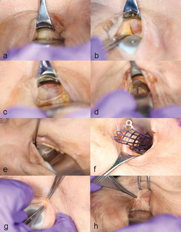
Fig. 1.

Cadaveric dissection illustrating the retrocaruncular approach to the medial orbit (looking toward the medial canthus). (a) The caruncle is retracted and protected with a Desmarres retractor and the globe protected with a malleable retractor. (b) After the retrocaruncular conjunctival incision, Horner muscle can be identified and left undisturbed. (c) Periosteum is incised just posterior to Horner muscle, and the bony medial orbital wall is visualized. (d) The anterior ethmoidal neurovascular bundle usually needs to be divided to expose the entire medial wall. (e) The posterior neurovascular bundle. (f) A small-sized prefabricated titanium plate is inserted into the orbit through an inferior fornix incision, which has been extended from the retroconjunctival incision. (g) Division of the caruncle in the transcaruncular approach. (h) The divided caruncle showing Horner muscle directly attached to the caruncle.
