Fig. 7.

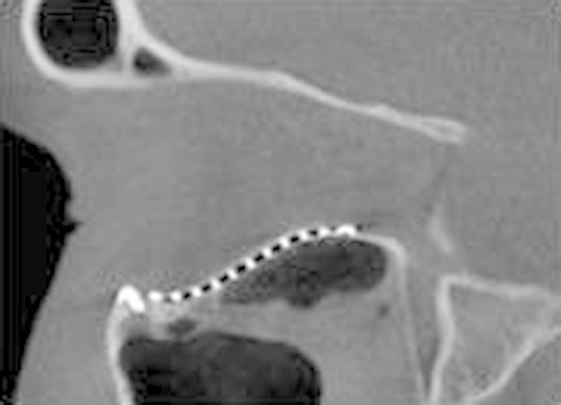
A sagittal section of an ICAT image demonstrating an orbital implant with optimal anatomical contours which restored the “key area” at the posterior equatorial bulge with the posterior aspect of the implant flush with the posterior ledge.

A sagittal section of an ICAT image demonstrating an orbital implant with optimal anatomical contours which restored the “key area” at the posterior equatorial bulge with the posterior aspect of the implant flush with the posterior ledge.