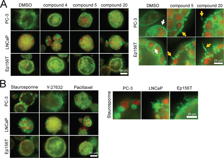
Fig 8. 3D morphologies of prostate spheroids, exposed to selected betulin derivatives and captured with a confocal microscope.
Actin cytoskeleton (filamentous or F-actin) is stained green (phalloidin), nuclei with a red dye (confocal microscope images, 40× objective, scale bar shown for each panel on the right lower corner).

