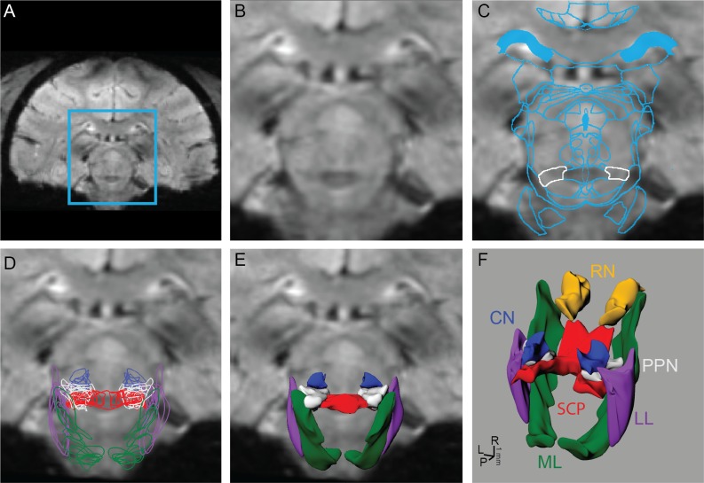
Fig 1. Process for reconstructing brainstem nuclei and fiber tracts in 3D from 7T MRI.
The brainstem region outlined in blue (A) was cropped (B) from each coronal 7T SWI MR image. (C) An affine deformation algorithm based on user-defined seed points was used to warp contours from a rhesus macaque brain atlas to the MRI of each subject. The PPN is outlined in white. (D) Algorithm-defined contours from nuclei and fiber tracts within brainstem were outlined on each slice and then (E, F) lofted to create surface renderings.

