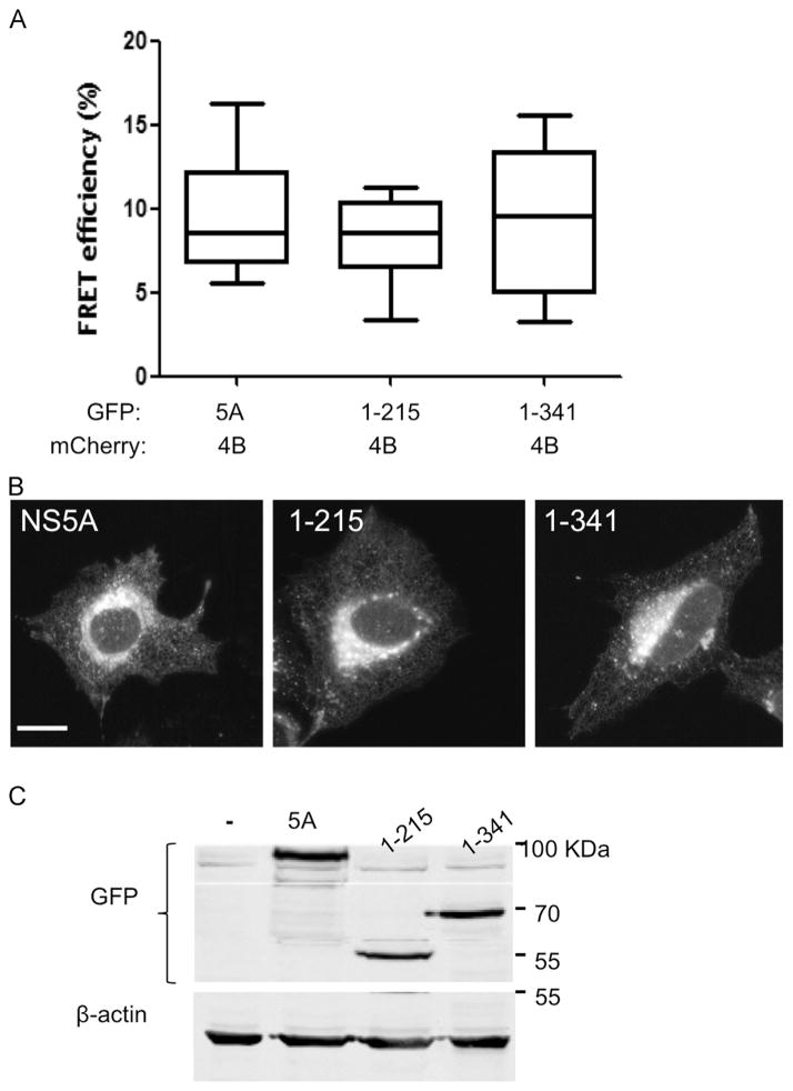
Fig. 6.
FRET analysis of the NS5A deletion mutants. A. U2OS cells were co-transfected with the indicated NS4B and NS5A combinations. Acceptor photo-bleaching FRET analysis was performed 24 h post-transfection (as described in Fig. 1). B. Subcellular localization of the NS5A deletion mutants. U2OS cells plated on coverslips were transfected with the indicated NS5A expressing constructs. Subcellular localization was determined using confocal microscopy 24 h post-transfection. C. Comparable expression of the NS5A deletion mutants was determined using western blot with anti-GFP antibodies. Monoclonal mouse anti-β-actin antibody was used as a loading control. Molecular mass markers are indicated on the right.

