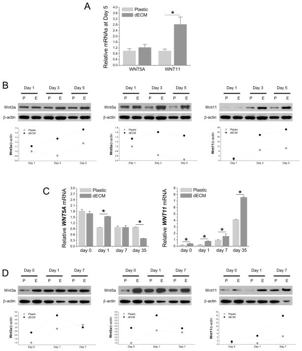
Fig. 6.
Wnt signals were actively involved in chondrogenesis of dECM expanded hSDSCs. Both real-time PCR (A, C) and Western blot (B, D) were used to evaluate the canonical Wnt signal (Wnt3a) and noncanonical Wnt signals (Wnt5a and Wnt11) in hSDSCs during cell expansion (A, B) and chondrogenic induction (C, D) at both mRNA and protein levels. The β-actin was used as an internal control. Data are shown as average ± SD for n=4. *p < 0.05 indicated a statistically significant difference. ImageJ software was used to quantify immunoblotting bands.

