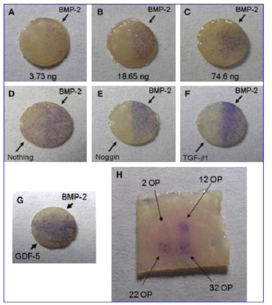
Figure 2.
ALP staining (blue) of C2C12 cells on 5 mm decellularized skin discs printed with bioactive factors resulting from (A-C) varying amounts of BMP-2 printed on the right halves of the scaffolds, (D-F) BMP-2 printed uniformly on the scaffolds with inhibitors printed on the left halves, (G) GDF-5 printed on the left halves, BMP-2 on the right halves, as well as (H) on a square piece with increasing number of BMP-2 overprints (OP). Adapted, with permission, from Cooper, et al. [179]. Copyright Mary Ann Liebert, Inc. 2010.

