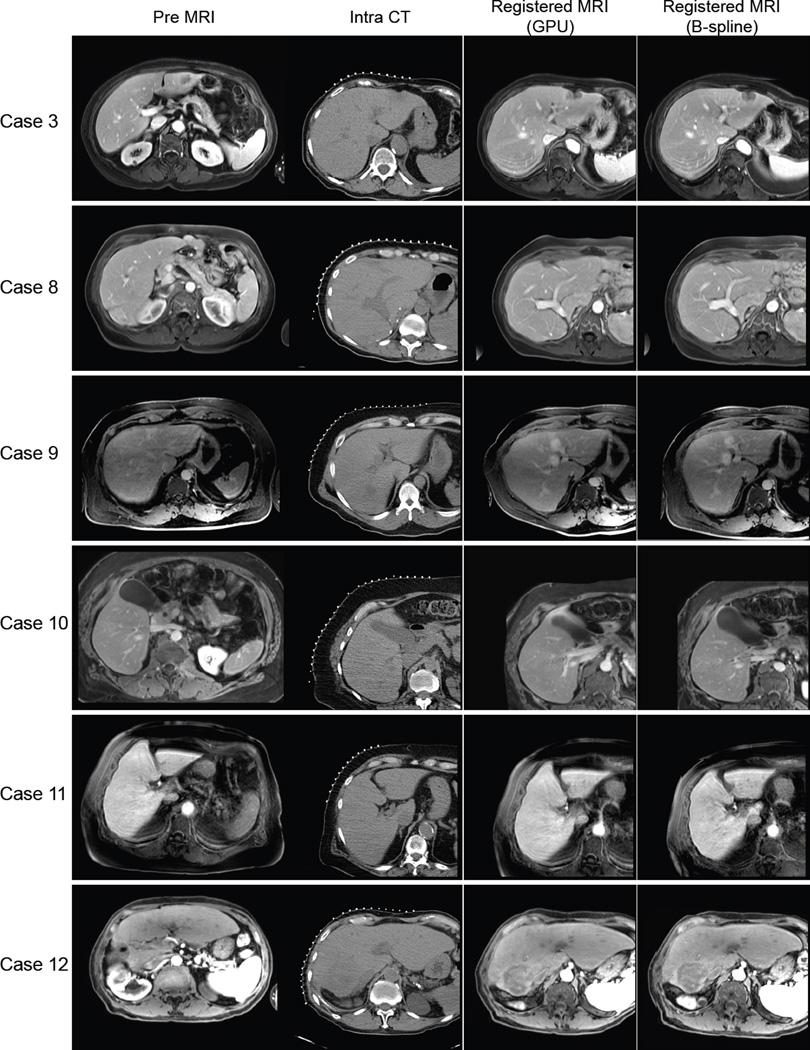
Figure 3.
The original preprocedural MRI, intraprocedural CT, and resampled MR images registered using the B-spline registration technique and the GPU-accelerated registration technique for representative cases are shown. Each row shows different procedures. The case numbers correspond to Table 2.

