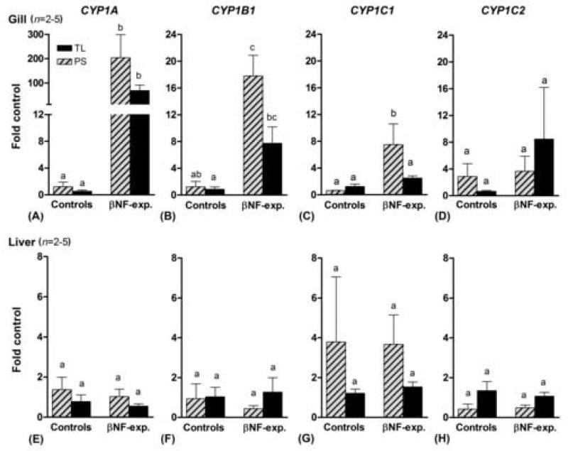
Fig 3.
CYP1A, CYP1B1, CYP1C1, and CYP1C2 expression in gills and liver of zebrafish TL (black bars) and PS (hatched bars) following exposure to β-naphthoflavone (1 μM) or the carrier (20 ppm acetone; mean ± SEM). For each CYP1 gene, basal and induced expression was calculated using the mean value of controls of both varieties as a calibrator. Differences between groups were determined by one-way ANOVA followed by Tukey's Multiple Comparison Test. A statistical difference between groups at p < 0.05 is indicated by differences in the letters above the bars.

