Figure 1.

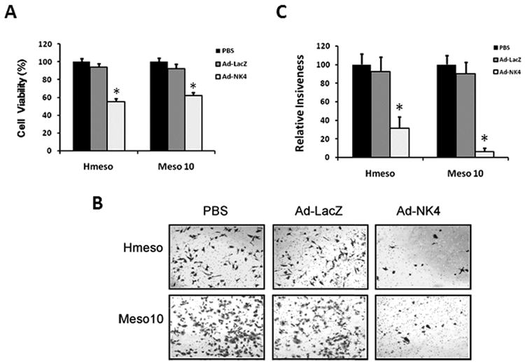
Expression of NK4 inhibits MM cell viability and invasiveness. (A) Inhibition of MM cell viability by Ad-NK4. MM cells Hmeso and Meso 10 were plated at a density of 5 × 10³ cells per well in 96-well tissue culture plates and grown in RPMI 1640 medium supplemented with 5% FBS and 10 ng/ml HGF. After overnight incubation, the cells were mock-infected or infected with Ad-LacZ or Ad-NK4. Cells were incubated for another 72 h, followed by MTT assay. Results are expressed as percent cell viability relative to the viability of control mock-infected cells (PBS). Each bar represents the mean value of five replicate wells from a representative experiment (n=3). *, p < 0.01 versus control. (B) Inhibitory effect of Ad-NK4 on invasiveness potential of MM cells in a Matrigel invasion assay. Hmeso and Meso 10 cells were mock-infected (PBS) or infected with Ad-LacZ or Ad-NK4 for 24 h. The following day, cells were seeded in the upper compartment of a Matrigel chamber in serum-free medium and allowed to invade overnight toward 10 ng/ml HGF, used as a chemoattractant, present in the lower compartment. Representative bright-field microscopic fields showing stained cells that invaded through the Matrigel. (C) Quantitative analysis of invasion assays. Ad-NK4 infection markedly decreased the average number of invading cells. Bars represent mean number of counted cells in five different microscope fields ± s.d. *, p < 0.01 compared to mock-infected cells.
