
Figure 4.
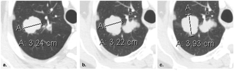
Failure to change measurement axis with changes in lesion orientation (51-year-old male with metastatic adenoid cystic carcinoma of the tongue base). Baseline contrast-enhanced computed tomography of the chest viewed at lung window settings (a) demonstrates a mass in the right lung, correctly measured along its axis. At 8-week (time point #2) follow-up imaging (b), the original measurement axis was incorrectly maintained, resulting in the underestimation of true lesion size. Shifting the measurement to the new long axis (c) correctly captures the interval lesion growth.