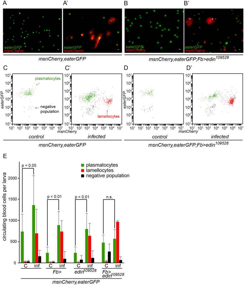
Fig 3. Quantification of hemocytes in edin RNAi larvae after a wasp infection.
(A-B) Hemocytes of infected larvae were bled 48–50 hours post-infection and visualized with the eaterGFP (green) and msnCherry (red) reporters. Uninfected controls contained only GFP-positive cells that corresponded to plasmatocytes (green). (A’ and B’) msnCherry expression was detected in the infected samples and this included lamellocytes (asterisks) and cells that express both eaterGFP and msnCherry indicating that they were undergoing lamellocyte transition. Lamellocytes were present also in the infected edin RNAi larvae suggesting that edin expression is not necessary for lamellocyte differentiation. Scale bars are 10 μm (C-E) Flow cytometry was carried out to quantify the amount of hemocytes in the unchallenged and the wasp infected edin RNAi larvae. (C = control, inf = infected)

