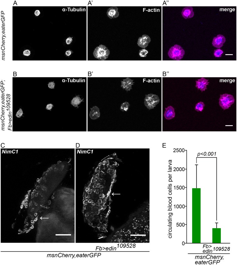
Fig 4. Edin expression in fat body is dispensable for normal hemocyte attachment to and spreading on glass and wasp eggs, but is necessary to increase blood cell numbers in circulation early after wasp infection.
(A-B”) Hemocytes from infected control larvae (msnCherry,eaterGFP, A-A”) and from infected larvae in which edin was knocked down in the fat body (msnCherry,eaterGFP;Fb>edin 109528, B-B”) spread normally on glass 14 hours after wasp infection despite knock down of edin in fat body. The spreading ability of hemocytes was assayed by staining α-Tubulin (blue) and F-actin (magenta). The size bar denotes 10 μm. (C and D). Wasp eggs from infected control larvae (msnCherry,eaterGFP, C) and from infected larvae in which edin was knocked down in the fat body (msnCherry,eaterGFP;Fb>edin 109528, D) were stained with the anti-plasmatocyte antibody NimC1. The wasp eggs were dissected 14 hours after parasitization and are still attached to the gut. Plasmatocytes spread normally on the eggs irrespective of edin RNAi in the fat body. Arrows denote examples of plasmatocytes spreading and adhering normally on the surface of the wasp egg. The scale bar depicts 50 μm. (E) Edin RNAi in the fat body (msnCherry,eaterGFP;Fb>edin 109528) reduced the number of circulating cells after wasp infection in comparison to control larvae (msnCherry,eaterGFP) 14 hours after infection. Circulating blood cell numbers were obtained with flow cytometry.

