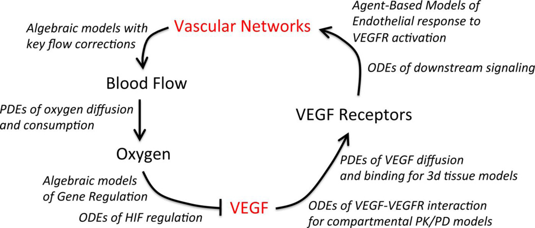
Figure 3. An example of vascular homeostasis and regulation by VEGF.
The many different computational model types employed to simulate the flow of information through the integrated multi-scale physiological models are indicated in italics. In general, models of in vivo pathology incorporate key elements of tissue physiology: vascular network geometry, blood flow, and/or oxygen distribution. Detailed models of molecular and cellular regulation, for example of the VEGF family, are often constructed and validated with in vitro experimental data, and then integrated into in vivo models and coupled to the other scales of regulation (Fig. 2) to predict the vascular remodeling and other physiological changes resulting from molecular perturbations (such as therapeutics). In diseases such as cancer, the homeostatic regulatory mechanisms can become non-functional or function in altered ways, leading to different vessel morphology than observed under physiological conditions.

