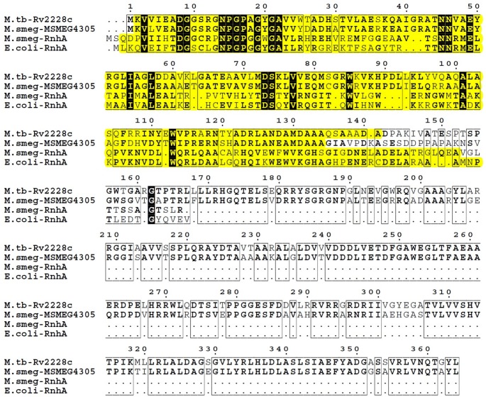
Fig 2. Comparison of protein sequences of RNases H type I.
Sequence alignment between RNases H type I of E. coli K12_MG1655 (RnhA), M. smegmatis mc2 155 (MSMEG4305 and RnhA) and M. tuberculosis H37Rv (Rv2228c) was performed using MultiAlin and visualized with ESPript 3.0. Highly similar or identical residues between protein sequences are written in bold. Identical residues across all analyzed sequences are shown in white on a black background. Similarities between protein sequences are marked by framing. The span of RNase H domains in each protein sequence, as defined by SMART, is highlighted in yellow.

