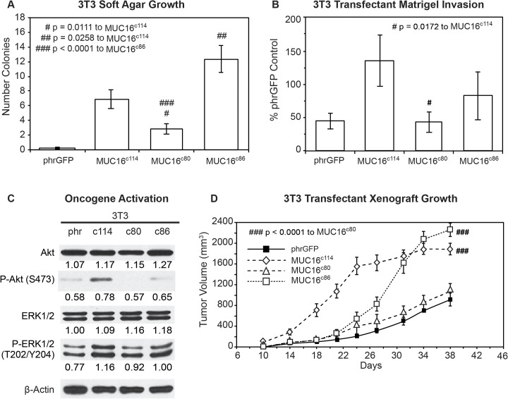
Fig 4. Effects of truncated MUC16c114 variants.
A) Soft agar growth. 3T3 transfectants expressing either internal or external domain portions of MUC16c114 (S1 Fig) were layered on soft agar, as described in the Material and Methods section. Colonies were counted and plotted. The data shown represent one of three experiments. Soft agar growth rates for MUC16c80 and MUC16c86 were significant (# p = 0.0111 and ## p = 0.0258, respectively) compared to MUC16c114, whereas a higher level of significance (###p<0.0001) was seen with MUC16c80 transfectant compared to MUC16c86. B) Matrigel invasion assay for 3T3 cell lines transfected with either phrGFP control vector or with MUC16 carboxy-terminal constructs. Each assay was performed two or more times in triplicate and counted by hand. MUC16c80 transfectant was significant (# p = 0.0172) compared to the MUC16c114 cell line. C) Effect of MUC16 expression on ERK/AKT signaling. Transfected 3T3 cells were examined for activation of the ERK/AKT signaling pathways. Phosphorylation of ERK1/2 (pT202/Y204) and AKT (S473) was increased following MUC16c114 transfection; however, the signals were lower with either the MUC16c80 or MUC16c86 constructs. β-Actin normalized densitometry quantification values are shown below each Western blot. D) MUC16-positive tumor growth in athymic nude mice. Two million tumor cells were introduced into the flank of 20 nu/nu mice, and the mice were observed for tumor formation. Tumors were measured by calipers twice weekly. The differences in mean tumor volume were significantly greater for mice bearing MUC16 ectodomain positive tumors. 3T3 MUC16c114 and 3T3 MUC16c86 transfectants were significantly different compared to MUC16c80 (### p<0.0001) and vector only animals.

